Identify the internal features of the heart.
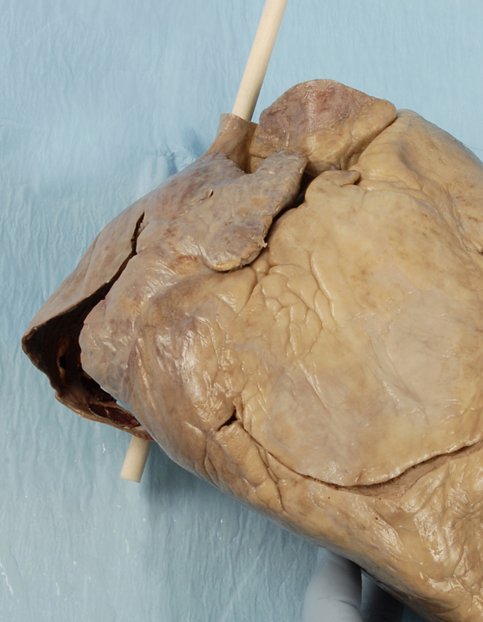
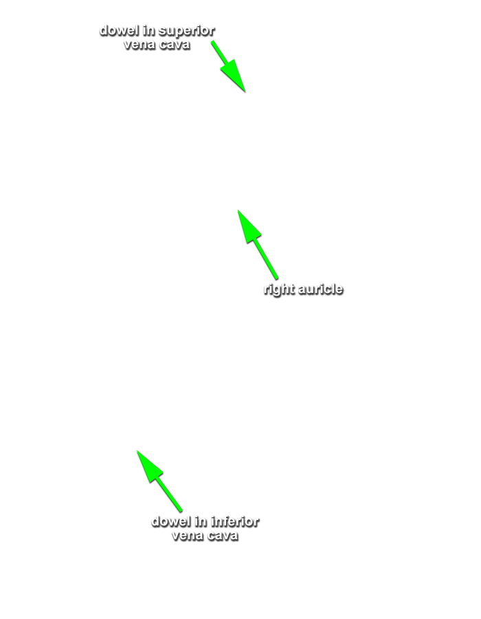
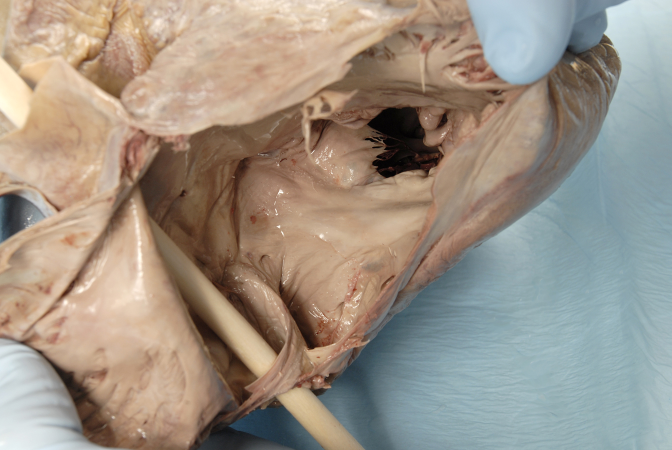
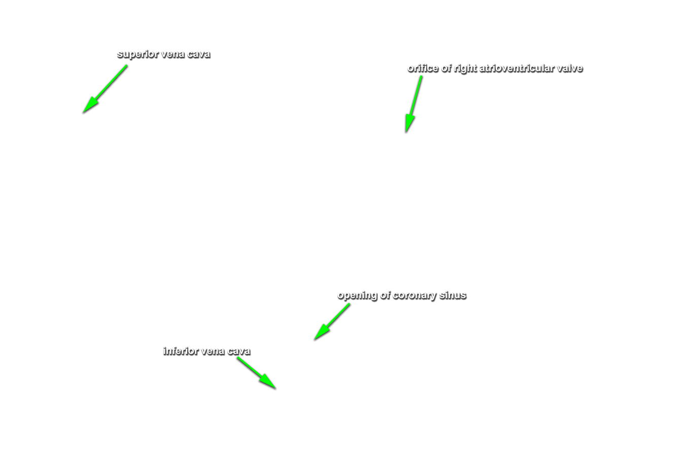
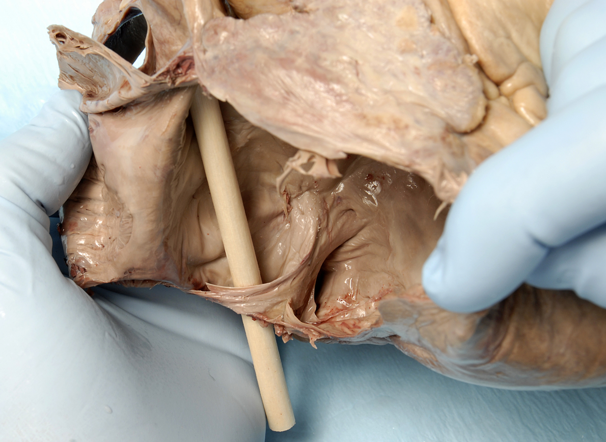
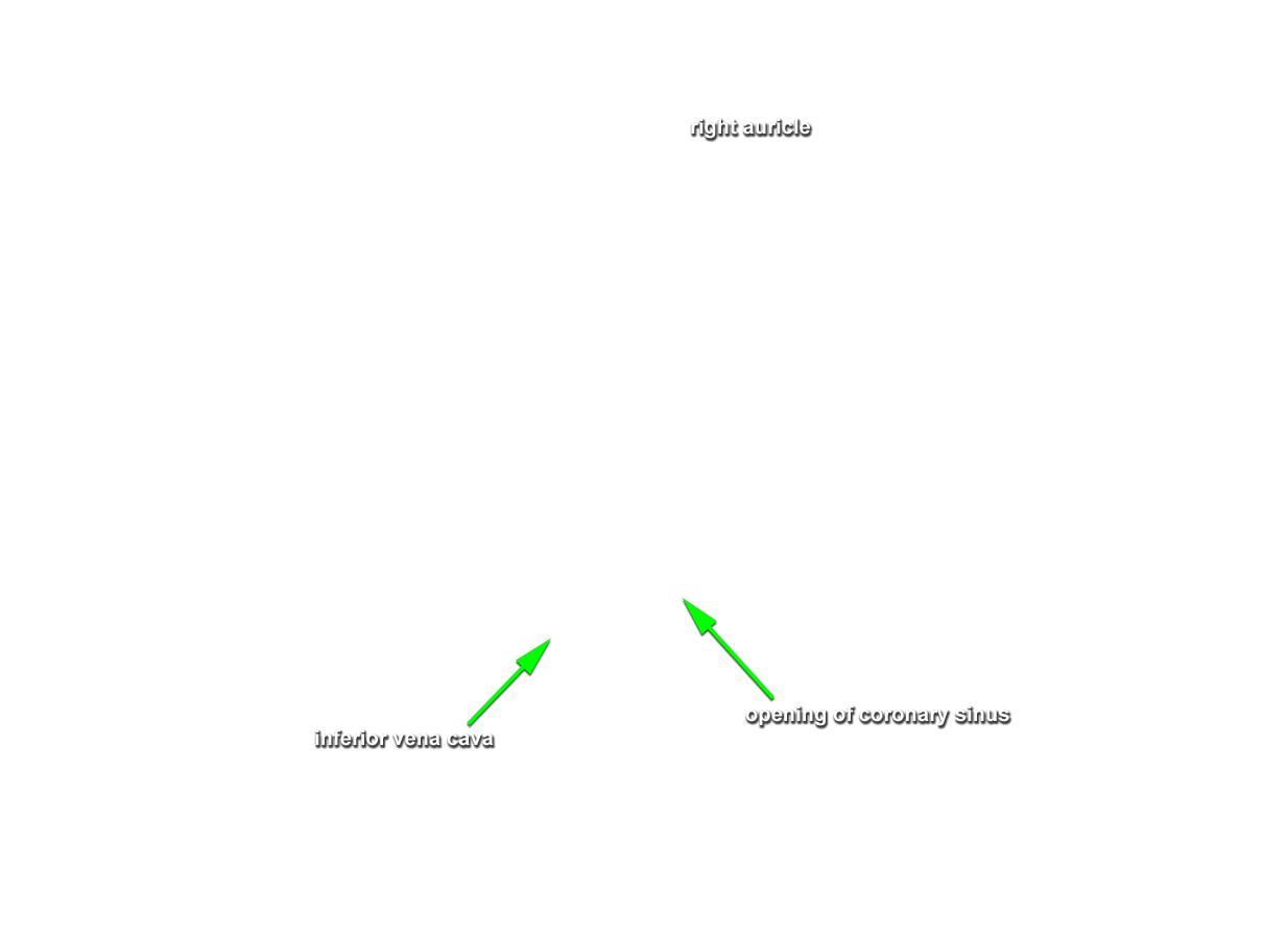
- Return to the superior vena cava and the inferior vena cava . (G 3.54A;N 217;Gl 9.11) Use your forceps to remove as much clotted blood as possible from the SVC, IVC and right atrium. Use pointed scissors to open the right atrium. Make a small cut in the tip of the auricle and then extend the incision along the superior border of the right atrium until you reach the SVC (but do not cut the SVC). Make a second incision along the medial (left) and inferior border of the right atrium until you reach the IVC (but do not cut the IVC). Fold the anterior wall of the atrium in the lateral (right) direction. Remove any remaining clotted blood and rinse the atrium with water.
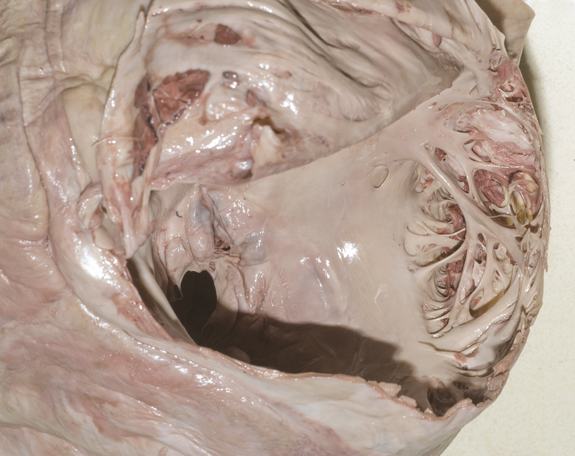
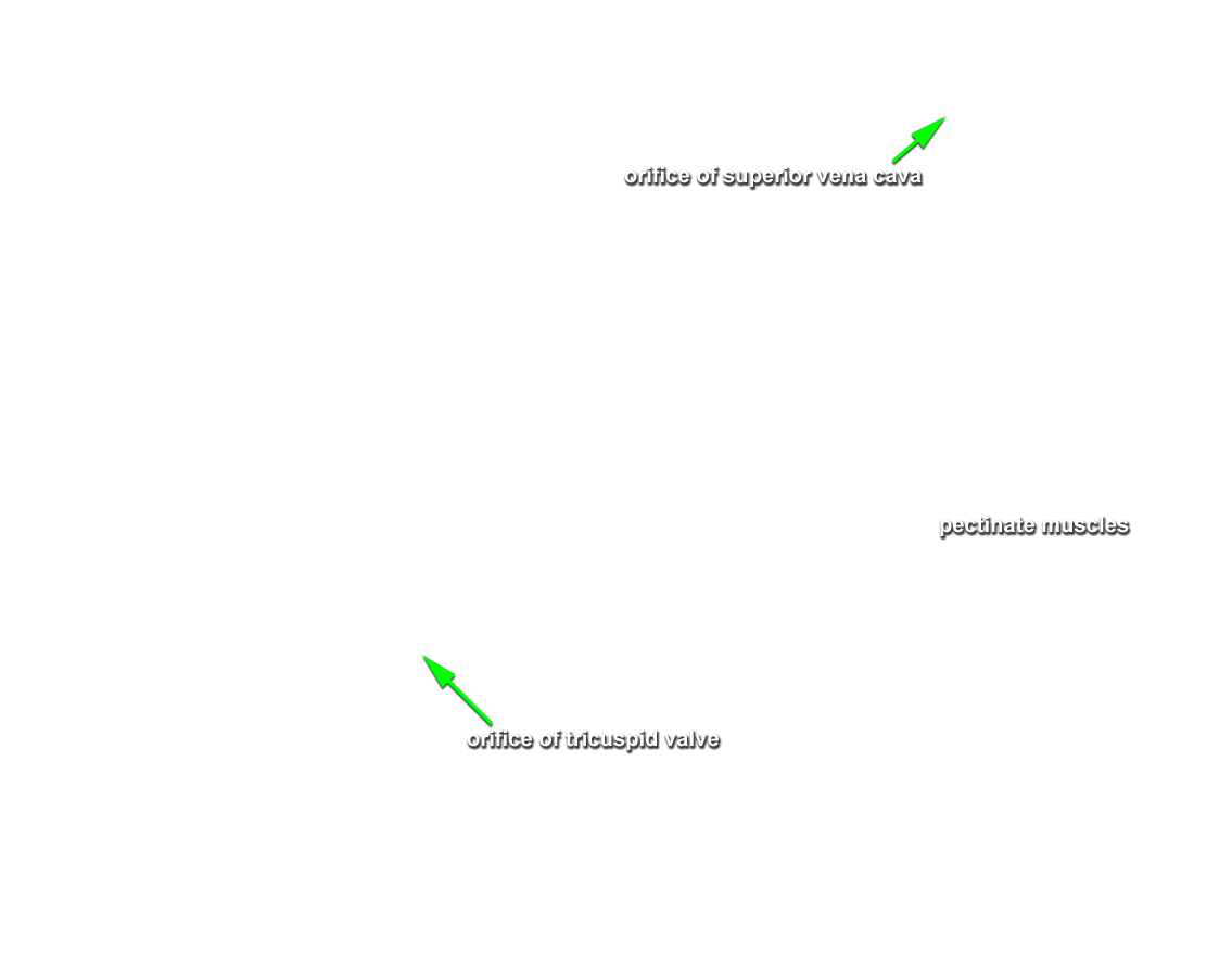
- Identify the
pectinate muscles forming the rough walled portion of the right atrium. Identify the
openings of the superior vena cava and the
inferior vena cava. Attempt to identify the valve of the IVC. Identify the
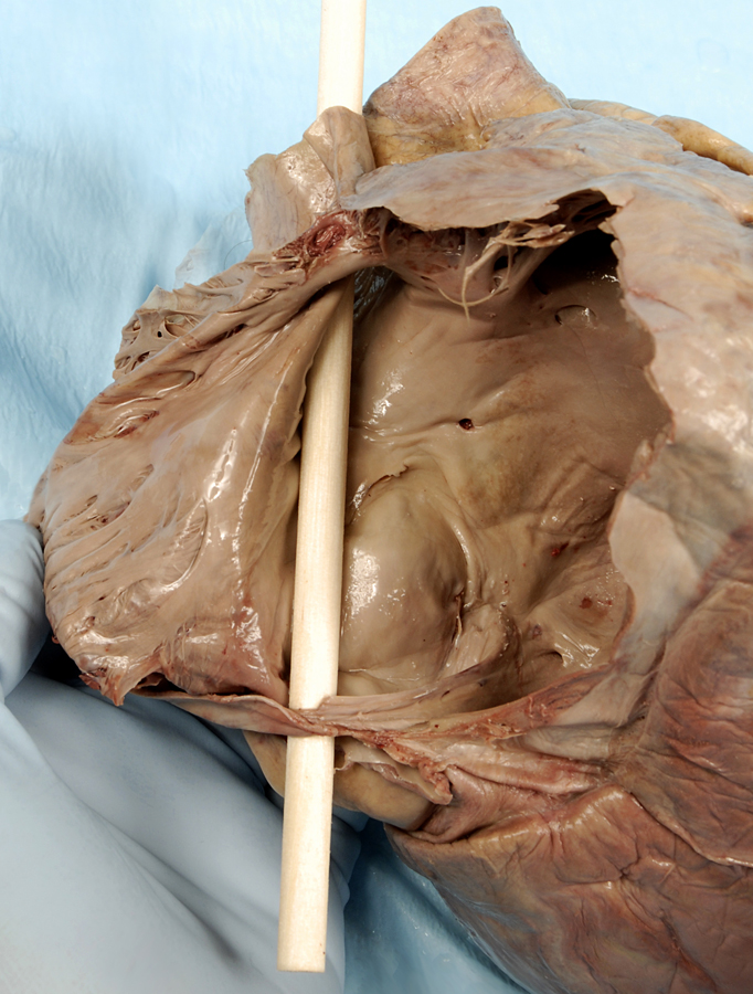
orifice of the right atrioventricular (tricuspid) valve. Identify the smooth walled
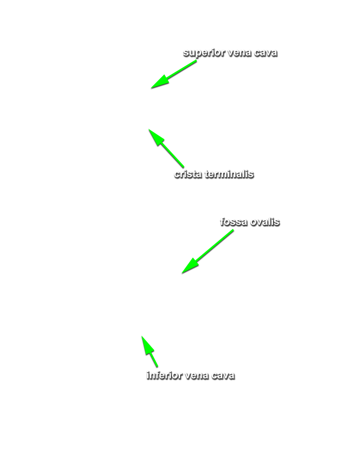
interatrial septum with the
fossa ovalis (oval fossa) and the
opening of the coronary sinus. Identify the
crista terminalis. This is a ridge of tissue where the rough and smooth walled portions of the right atrium meet (hopefully where you folded back the atrial wall. (G 3.54;N 217;Gl 9.12B) The S-A node is positioned where the crista terminalis meets the base of the SVC. The A-V node is located in the interatrial wall immediately superior to the opening of the coronary sinus.
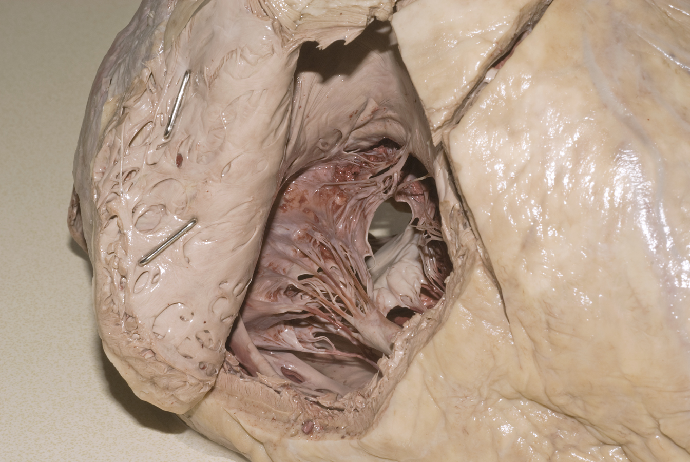
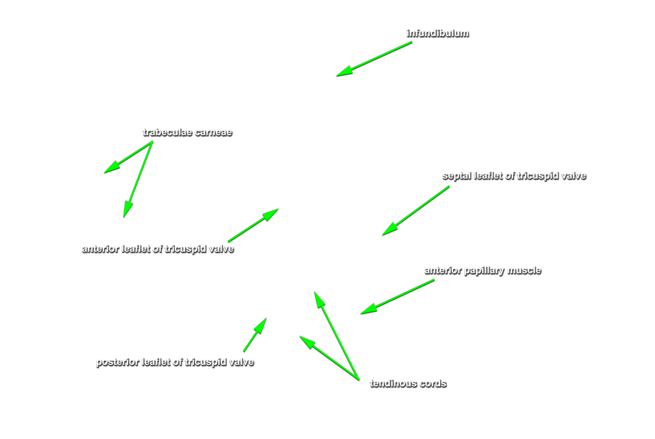
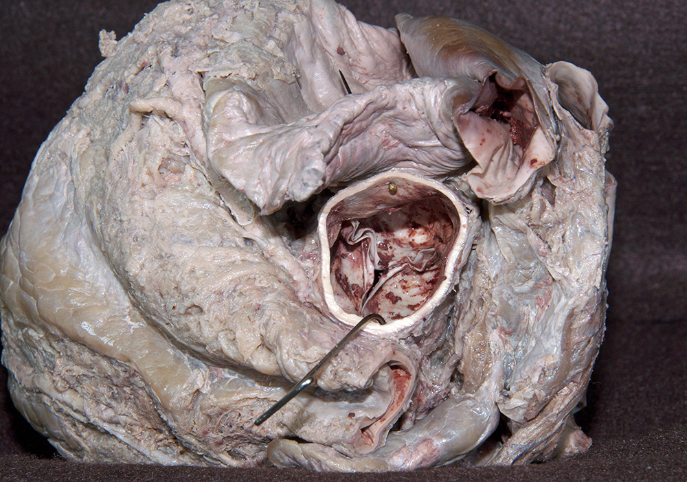
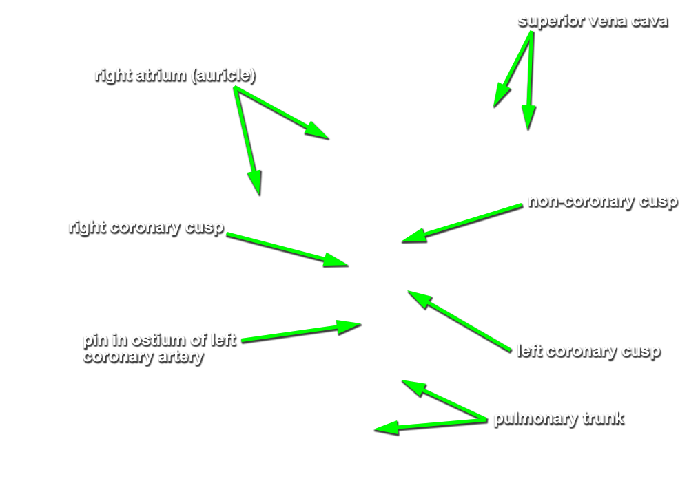
- Return to the pulmonary trunk. Identify the cusps of the pulmonary valve from the superior view. (G 3.58;N 219;Gl 9.13) Use your scissors to make an incision between the anterior and right cusps of the valve. Extend the incision through the anterior wall of the right ventricle towards the apex of the heart. Open the right ventricle with your fingers and remove the clotted blood. Rinse the right ventricle with water. Identify the muscular ventricular wall with the trabeculae carnae. (G 3.55;N 217;Gl 9.12A) If the ventricular wall is too thick and you cannot identify structures within the right ventricle, then remove a portion of the anterior wall as demonstrated in Grant's Figure 3.55. Identify the cusps, chordae tendinae (tendinous cords) and papillary muscles of the tricuspid valve. Attempt to identify the septomarginal trabecula extending between the base of the anterior papillary muscle and the interventricular septum. Identify the cusps of the pulmonary valve from the inferior view and the associated conus arteriosus (smooth walled region immediately inferior to the pulmonary valve).
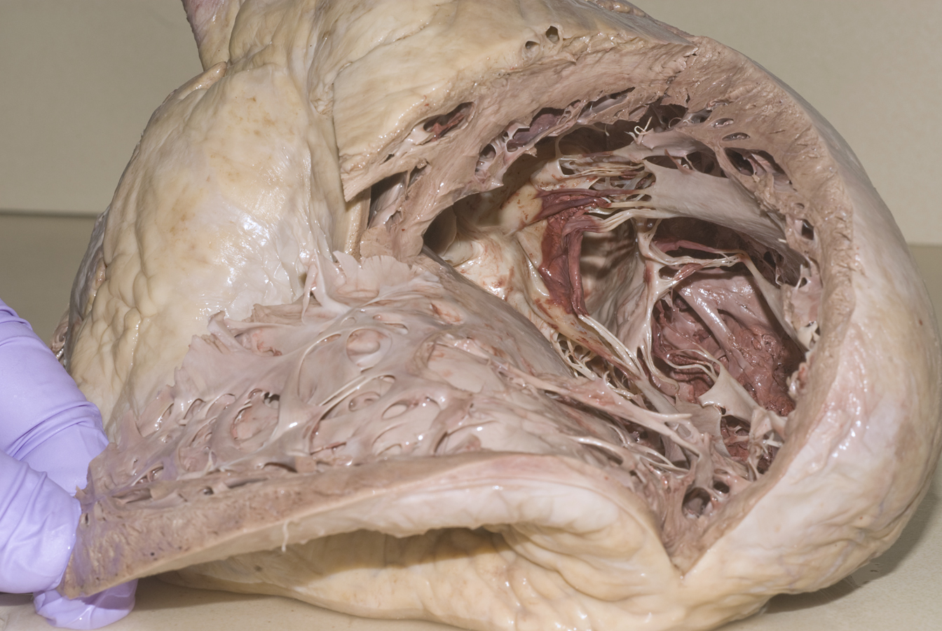
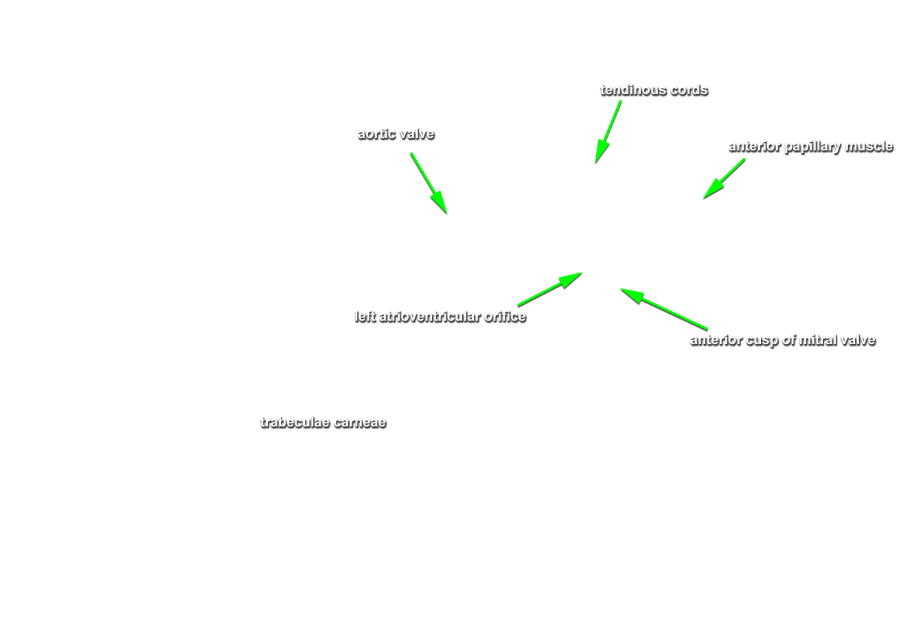
- Return to the ascending aorta. Identify the cusps of the aortic valve from the superior view. (G 3.58;N 219;Gl 9.13) Identify the orifices of the right and left coronary arteries. Use scissors to split the ascending aorta between the right and left cusps. Extend the incision through the ventricular wall to the apex of the heart. (G 3.57;N 218;Gl 9.12C) You will need to cut the anterior interventricular artery and the great cardiac vein during this procedure. Use your fingers to open the ventricle and remove the clotted blood. Rinse the ventricle with water. Identify the interventricular septum, and trabeculae carnae. Compare the thickness of the left and right ventricular walls. Identify the cusps, tendinous cords and papillary muscles of the bicuspid (mitral) valve.