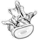
Figure 1-5
Vertebral disc attached to superior end of a lumbar vertebrae. Superior view.
Vertebral disc attached to superior end of a lumbar vertebrae. Superior view.
The numerous bones and their articulations combined with great strength requirement, result in a bony vertebral column connected throughout its length by intervertebral discs and numerous sets of strong ligaments. The ligaments in cooperation with muscles, limit movement and contribute to maintaining the integrity of the column during physical activity.
Intervertebral discs are fibroelastic cartilage plates firmly attached to adjacent vertebral bodies. Each disc is constructed of outer concentric cartilaginous rings called the annulus fibrosus. The rings of the annulus fibrosus resemble the growth rings of a tree. The annulus fibrosus surrounds a central jelly-like mass, the nucleus pulposus. This arrangement makes each disc a very tough shock absorber. (Figure 1-5)
The anterior longitudinal ligament runs vertically along the anterior side of the vertebral bodies and is continuous from the sacrum to the skull. This ligament limits hyperextension.
The posterior longitudinal ligament runs vertically along the posterior side of the vertebral bodies and is continuous from the sacrum to the skull. It limits hyperflexion. The posterior longitudinal ligament lies in the vertebral canal, anterior to the spinal cord.
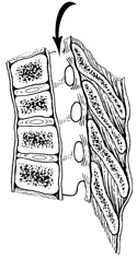
The yellow ligamenta flava connects adjacent vertebral lamina and helps form the roof of the vertebral canal. It may also limit lateral flexion. (Figure 1-6)
The interspinal ligament connects adjacent vertebral spinous processes, while the continuous supraspinal ligament runs immediately posterior to the vertebral spinous processes from the sacrum to the skull. Both of these ligaments limit hyperflexion. (Figure 1-6)
In the cervical region, the interspinal and supraspinal ligaments appear to enlarge and combine to form the nuchal ligament. This large sheet of midsagittal connective tissue serves as a place for many muscles to attach as well as a supportive structure for the head.