Figure 1-12
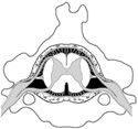
Spinal meninges at level C3.
Spinal meninges at level C3.
The spinal cord and brain are enclosed by three protective connective tissue membranes called meninges. The spinal meninges are continuous with the cranial meninges but exhibit some notable differences.
Characteristics of spinal meninges:
Clinically important, the distal end of the spinal cord and meninges are the site for diagnostic and anesthetic procedures. In the adult, the spinal cord ends at bony level L2. (Figures 1-13 and 1-14)
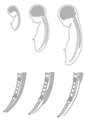
Three months prior to birth, the bony vertebral column, spinal cord and meninges are the same length. Development brings about differential growth rate. The bony vertebral column and canal, with distally attached meninges, grow faster than the spinal cord. The cord appears to "shrink" within the vertebral canal, however, it is still growing but the relatively slower rate of growth creates the illusion of shrinkage. Thus, at maturity the conus medullaris, the end of the spinal cord, ends at bony level L2.
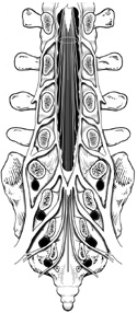
The dura mater and arachnoid, however, grow with the bony column to the S2 level. The pia mater retains its adherence to the cord tissue except for a small strand called the filum terminale attached to the coccygeal vertebrae anchoring the cord inferiorly. (Figure 1-13)
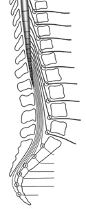
This differential growth rate leaves a large subarachnoid space between the conus medullaris (L2) and the distal ends of the arachnoid and dura mater (S2), called the lumbar cistern. The collection of numerous spinal nerve roots passing through the lumbar cistern creates the appearance of a horse's tail, the cauda equina. (Figure 1-14)
*Question: If the spinal cord ends at bony level L2, how do the lower lumbar and sacral spinal nerves get to their appropriate intervertebral foramina?
*Hint: Spinal nerve L5 exits the vertebral column below the fifth lumbar vertebra. The spinal nerve S2 exits below the second bony segment of the sacrum.
*Answer: Differential growth rate causes the lower lumbar and sacral vertebrae to grow distally pulling the spinal nerves with them. After emerging from the spinal cord, the nerves travel inferior in the lumbar cistern to their appropriate intervertebral foramina located inferior to L2. (Figure 1-15)
To obtain CSF, a syringe is inserted into the lumbar cistern (subarachnoid space) below L2 and above S2.
The spinal cord is superior to the needle entry and will not be injured. The spinal nerves in the cauda equina area will move out of the way like spaghetti in the path of a pointed knife.