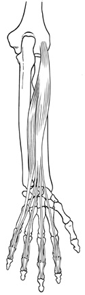
Figure 12-3
Superficial extensors of the anterior forearm.
Superficial extensors of the anterior forearm.
Locate the lateral epicondyle and look for these extensor muscles as they pass to the wrist or the digits. Take note of the position of the forearm to see if it is pronated or supinated. Our description is of the supinated forearm viewed from a posterior aspect.
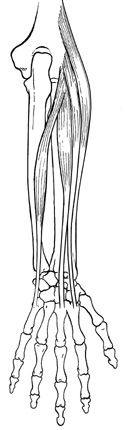
The most superficial and laterally placed muscle in this group is the brachioradialis. It runs from the lateral supracondylar ridge to the styloid process of the radius. At first glance, this muscle has all the characteristics of an elbow extensor, since it's innervated by the radial nerve and lies on the lateral side of the forearm. The brachioradialis, however, is really an elbow flexor in the mid-prone position.
Moving medially, you will encounter the two radial extensors of the wrist, the more lateral, short-bellied and long-tendoned extensor carpi radialis longus, followed by the more medial extensor carpi radialis brevis. These muscles can easily be identified if you see their distal attachment. The extensor carpi radialis longus inserts on the second metacarpal, the extensor carpi radialis brevis inserts on the third metacarpal.
The largest of the superficial posterior forearm muscles, and closest to the midline of the forearm, is the extensor digitorum. This muscle arises from the lateral epicondyle and sends tendons to the phalanges of digits 2-5. The extensor digiti minimi attaches distally to the fifth digit and is really a specialized part of the extensor digitorum. Locate both of these muscles, taking extra care to observe their distal attachments, the middle and distal phalanges of digits 2 through 5.
Note how the tendons of these muscles seem to flare out and get thinner as they cross over both the M-P joint and the proximal phalanx of digits 2-5. Can you also see two "lateral bands" of the dorsal digital expansion break away and stay on the outer edge of the proximal interphalangeal joints? They should cross the proximal I-P joint and then reunite over the distal I-P joint, inserting on the distal phalanx of digits 2-5. Notice also a "central band" continuing over the posterior surface of the proximal I-P joint to insert on the dorsum of the middle phalanx. This complex dorsal digital expansion mechanism allows the extensor digitorum to extend both the proximal and distal I-P joints. It also provides an effective point of distal attachment for the lumbricales and interossei.
The extensor carpi ulnaris is the last extensor muscle in the superficial group. It's the most medially placed muscle of all the superficial posterior group, attaching distally to the fifth metacarpal.