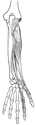
Figure 12-5
Deep extensors of the anterior forearm.
Deep extensors of the anterior forearm.
This is a mixed group of muscles. Deeply placed, the supinator arises off the lateral epicondyle of the humerus and inserts on the radius. In locating the distal end of the pronator teres, you also find the distal end of the supinator.
The
extensor indicis proprius
 arises from the distal ulna and runs distally to join the tendons of the
extensor digitorum (ED).
Look for this muscle's tendon as it leaves the
ED
tendons and goes deep into the posterior and distal forearm.
arises from the distal ulna and runs distally to join the tendons of the
extensor digitorum (ED).
Look for this muscle's tendon as it leaves the
ED
tendons and goes deep into the posterior and distal forearm.
The remainder of these deep extensor muscles attach proximally on the distal half of the radius and the interosseus membrane and run to the thumb. For this reason they appear to emanate from the depths of the distal posterior forearm and are sometimes called "outcropping muscles."
The tendons of these three "outcroppers" appear on the lateral inferior portion of the forearm. Moving from lateral to medial in the anatomical position or from the radial to the ulnar side of the back of the hand, you can first see the tendons of the abductor pollicis longus, inserting into the first metacarpal, followed closely by the extensor pollicis brevis inserting into the base of the proximal phalanx of the thumb. The interval between these two muscles and the next muscle, the extensor pollicis longus, which inserts onto the distal phalanx of the thumb, is the "anatomical snuffbox." One can take a pulse here as the radial artery runs across the floor of this "snuffbox."
Again if you're lucky, you will find the superficial branch of the radial nerve. This nerve is the terminal end of the radial nerve and runs distally from the common origin of the extensor group on the lateral or radial side of the forearm. This is a cutaneous nerve to the posterior - radial side of the hand.