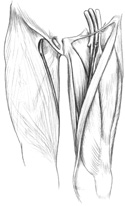
Figure 15-1
Major structures of the anterior thigh.
Major structures of the anterior thigh.
If the abdomen and pelvis have been dissected, look into the pelvic cavity and find the external iliac artery and vein. These are the blood supply and return routes of the lower limb. When these vessels pass directly under the inguinal ligament, which is really the inferior border of the external abdominal oblique, they become the femoral artery and vein. Find the femoral artery and vein just inferior to the inguinal ligament.
Immediately lateral to the femoral artery, look for the femoral nerve. It soon splits into several branches to serve the anterior thigh region. (Figure 15-1)
In this same general area, see if you can find the largest branch of the femoral artery the profunda femoris artery. This artery leaves the femoral artery laterally, slightly inferior to the inguinal ligament and is posterior and medial to the branching femoral nerve.
The profunda femoris artery soon gives rise to a lateral and medial circumflex femoral artery that leave the profunda femoris according to their name, and encircle the proximal end of the femur. The lateral circumflex also gives rise to a descending branch, which supplies the anterior thigh region.
The profunda femoris continues inferiorly, giving rise to several perforating branches that "perforate" their way to the posterior thigh.
The femoral artery and vein continue inferiorly under the sartorius muscle, which runs from the anterior superior iliac spine to the medial proximal tibia. This subsartorial canal is frequently called the adductor canal because the artery and vein lie on top of (anterior to) the adductor muscles.
Near the distal and medial end of the thigh, follow the femoral artery and vein as they disappear through the adductor magnus muscle by way of the adductor hiatus. They become the popliteal artery and vein on the posterior surface of the thigh in the popliteal fossa, or back of the knee.
The rectus femoris is the most superficial of the anterior thigh muscles, crossing the hip and originating from the anterior inferior iliac spine (Figures 15-1, 15-2).
Now identify the three vasti muscles. The vastus lateralis lies on the lateral side of the thigh.
The vastus medialis covers the medial aspect of the thigh. The vastus intermedius is positioned between the vastus medialis and lateralis.
These four muscles are collectively called the quadriceps femoris.
The muscles of the anterior thigh, all except the sartorius, attach distally to the tibial tuberosity by way of the patellar ligament (tendon). This "ligament" is nothing more then the continuation of the quadriceps tendon after it has passed over and around the patella.
The patella is a sesamoid bone, developed within the quadriceps tendon.