Figure 2-6
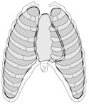
Anterior view of the pleural sacs and mediastinum within the thoracic cavity.
Anterior view of the pleural sacs and mediastinum within the thoracic cavity.
The thoracic cavity contains two pleural sacs and the mediastinum. The mediastinum is the space between the two pleural sacs. It contains the heart, aorta, azygous veins, trachea, esophagus, vagus nerves, sympathetic nerve trunks and other important structures. (Figure 2-6)
The two pleural sacs occupy the lateral portions of the thoracic cavity. During development the lungs grow into the pleural sacs much like pushing a fist into a balloon. This process completely covers the lungs with a visceral layer of pleura while the inside wall of the thorax is lined by a parietal layer of pleura.
Adult lungs are so large they take up most of the space in the balloon. The inside of the balloon (pleural cavity) is a thin space containing only a small amount of lubricating serous fluid. The presence of serous fluid creates surface tension between the two pleural layers. The parietal pleura is pulled away from the visceral pleura during inspiration by the expansion of the thoracic wall to which it is attached. The visceral pleura, adherent to the lungs, is attracted to the parietal pleura by the surface tension between the two layers. The resulting expansion increases the volume of the lungs.
The pleural cavities are two separately enclosed spaces except for an isthmus through which the airways and blood vessels enter and leave. This area of attachment to the mediastinum is called the root or hilus of the lung. (Figure 2-7) Here the visceral pleura is continuous with the parietal pleura.