Heart, Pericardium and Great Vessels
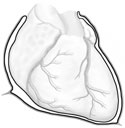
The heart and the great vessels, passing to and from it, lie in the mediastinum. They are enclosed
in a pericardial sac. This pericardium is composed of an outer single-layered, non-serous fibrous
pericardium, and an inner double-layered serous pericardium. The outer, fibrous pericardium is
attached to the diaphragm and sternum and helps anchor the heart. The inner pericardial sac is very
similar to the pleural sacs surrounding the lungs; the heart has grown into this sac so there
exists a visceral layer lying right on the tissue of the myocardium, and an outer parietal layer.
Like the pleural sacs, the pericardial cavity is a potential space between the two serous layers
and contains only a small amount of lubricating serous fluid; of great importance to the
continuously contracting heart. (Figure 2-9)
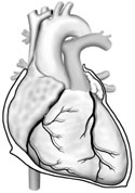
Eight vessels running to and from the heart appear to pierce the pericardium. They are the aorta
and pulmonary artery, the superior and inferior vena cava, and four pulmonary veins. (Figure 2-10)
When the heart is lying in its natural position, the right ventricle makes up the largest part of
the anterior surface and inferior border. The right border consists of the right atrium, while the
left border and apex of the heart is formed by the left ventricle.
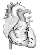
The myocardium is supplied with arterial blood by two sets of coronary arteries: a right and left.
Both arise from the ascending aorta. (Figure 2-11) Most of the coronary veins empty into a
coronary sinus which, in turn empties into the right atrium. (Figure 2-17)
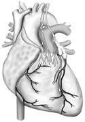
The heart can beat without any CNS nerve supply, however, it has sensory and motor innervation via
cranial nerve X. Parasympathetic and sympathetic fibers reach the cardiac plexus of the heart by
way of the superior and inferior cardiac nerves. (Figure 2-12)
The heart is divided into two parts to facilitate understanding its function:
-
The right heart receives O2 depleted blood from the body and pumps it to the lungs for
re-oxygenation, (the pulmonary circulation).
-
The left heart receives oxygenated blood from the lungs and pumps it back to all parts of the
body, (caval circulation).
The right heart has two chambers, the right atrium and right ventricle. The two chambers
communicate through the atrioventricular or tricuspid valve. The right atrium receives blood from
the superior and inferior vena cavae and pumps it to the right ventricle through the tricuspid
valve. The right ventricle pumps the blood to the pulmonary artery through the pulmonary semilunar
valve.
The left heart also has two chambers which communicate via the atrioventricular (A-V) valve.
Oxygenated pulmonary blood is received by the left atrium from the lungs. The blood is pumped to
the left ventricle through the left A-V valve. The left ventricle pumps blood to the aorta through
the aortic semilunar valve, the first step in its distribution to all parts of the body.
The extraordinary, synchronized function of all parts of the heart in providing a lifetime of
tireless service is one of nature's outstanding achievements.