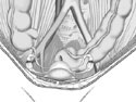
Figure 4-1
Female pelvic cavity. Anterior superior view.
Female pelvic cavity. Anterior superior view.
The female pelvic cavity contents include (from anterior to posterior) the urinary bladder, the uterus and the rectum.
Laterally we find the uterine tubes and ovaries. From a superior view, the two external iliac arteries form a horseshoe around these organs (Figure 4-1).
The urinary bladder, lodged tight against the posterior aspect of the pubic bone, receives urine from two ureters that have descended from the kidneys. The urinary bladder empties through the pelvic diaphragm to the exterior by way of a short separate tube, the urethra.
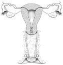
The female reproductive organs include four main components. First, the ovaries - organs that actually produce the genetic copy of the owner (the mature egg or ovum). Next are the uterine tubes, which not only transport the ovum from the ovary to the uterus, but also provide the usual sight of fertilization. The third element of the female reproductive system is the uterus. The uterus provides a nurturing home for the developing embryo to exist, as well as playing a large part in the birth process. The vagina is the fourth organ that serves a dual role: first as a route for the fetus to emerge into the world, and second, as an organ of sexual interaction (Figure 4-2).
During fetal development, the ovaries, anchored to the anterior abdominal wall by a connective tissue "ligament" called the gubernaculum, descend a short distance from their site of origin. The gubernaculum will eventually become attached to the uterus and form the ovarian ligament running from the ovary to the uterus. This union also forms the round ligament of the uterus, which runs from the uterus to the labia majora. The round ligament of the uterus passes through the inguinal canal on its way to the labia majora.
The ovaries are not directly attached to the end of the uterine tubes, but are completely surrounded by peritoneum and thus physically separated from the opening of the uterine tube.
The uterine tubes extend from the body of the uterus laterally, and open into the peritoneal cavity. The ends of the tubes have finger-like projections called fimbria that move and help guide the ovum into the uterine tube itself.
The uterus is the most powerful muscle in either the male or female; during labor contractions, it exerts 600 lb./square inch! This pear-shaped hollow organ has a main superior portion, the body of the uterus, and a smaller inferior neck-like part, appropriately called the cervix (neck) of the uterus. The cervix acts like a sphincter for the rest of the uterus during pregnancy and childbirth.
The vagina serves not only as the birth canal or "highway" from the uterus to the outside world, but also as the female organ of copulation. It receives the male organ of copulation, the penis and captures sperm cells during intercourse.
The rectum is the continuation of the sigmoid colon. It also pierces the pelvic diaphragm to empty its contents outside the body.
All of the organs of the pelvic cavity are only partially covered by peritoneum. Remember, this organ-peritoneal relationship is referred to as retro-peritoneal.
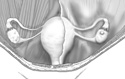
Even though the reproductive organs are considered to be retro-peritoneal, they have pushed substantially into the peritoneal sac, so much so that the uterus and its uterine tubes appear to be draped with peritoneum. This resembles someone standing upright with their arms outstretched, and a sheet draped over the top of them (Figure 4-3).
The folds of peritoneum connecting organ to organ, or organ to abdominal wall, are called "ligaments." With this in mind, the double layers of peritoneum hanging from the uterine tubes are called the broad ligaments. The layers of peritoneum sandwich uterine tubes, ovaries, ovarian blood vessels, ovarian ligament and round ligament of the uterus. The continuation of the broad ligament laterally to the body wall, is the suspensory ligament of the ovary and contains the ovarian vessels and the ureter.
The folds and layers of peritoneum anchor the reproductive organs to the body wall and provide avenues for vessels and nerves to follow.
Interestingly, because the ovaries and uterine tubes are not directly connected, the only way mature ova can reach the opening of the uterine tubes is to rupture the peritoneum covering the ovary (ouch!) leaving it in the peritoneal cavity near the fimbria of the uterine tube opening. The ova then enters the ostium (opening) of the uterine tube. There is, of course, the possibility that the egg can get lost on this perilous trip. The lost egg can be fertilized and partially develop in the peritoneal cavity as an ectopic pregnancy.
There is no comparable opening to the peritoneal cavity from the reproductive tract of the male.