Figure 7-12
Hemisected brain (right half).
Hemisected brain (right half).
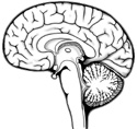
Before attempting to identify parts of the last primary brain structure, the brain stem, diverge for a moment and look at the ventricular system of a hemisected brain. Knowledge of this cavernous system in the brain will help you locate and identify the components of the brain stem. (Figure 7-12)
Looking at the medial surface of the cerebral hemisphere reveals a lateral ventricle conforming to the shape of the hemisphere. Depending on the way the brain was cut, you may see a thin partition separating the two lateral ventricles, the septum pellucidum.
Each lateral ventricle communicates with the third ventricle through an
interventricular foramen
 ,
located near the anterior aspect of each lateral ventricle. The roof of the lateral ventricles is formed by the
corpus callosum.
,
located near the anterior aspect of each lateral ventricle. The roof of the lateral ventricles is formed by the
corpus callosum.
Follow the interventricular foramina inferiorly to the
third ventricle.
This narrow cavity lies in the midline between the paired diencephalon (superior most part of brain stem) and is shaped like a doughnut; the center of the ventricle is occluded by a connecting bridge of nerve tissue. The third ventricle tapers inferiorly and forms the canal of the midbrain, the
cerebral aqueduct
, which communicates with the
fourth ventricle.
The pyramid shaped
fourth ventricle
lies anterior to the cerebellum, and posterior to the pons and medulla. This ventricle continues inferiorly as the central canal of the spinal cord and communicates with the subarachnoid space via three foramina in its roof. In any of the ventricles see if you can find a modified plexus of capillaries, the
choroid plexus
, which is responsible for the production of cerebrospinal fluid.