Figure 7-13
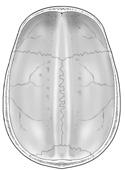
Interior skull cap.
Interior skull cap.
Now focus on the meningeal layer, the dura mater. Using a dura mater skull "cap" that has been removed from a cadaver, orient yourself by locating the superior and anterior aspects of this cap. On the superficial aspect of the lateral side, you should be able to identify the
middle meningeal vessels
 as they spread out to supply the dura with arterial blood. You can now appreciate how, if ruptured, these vessels would create an epidural hematoma (blood clot). (Figure 7-13)
Identify the two prominent inner dural infoldings, the mid-sagittal falx cerebri and the transverse tentorium cerebelli. (Figures 7-6)
Can you see any of the bony cranial attachments of these infoldings? Can you visualize the dural cap as it would be in the cranial vault? Now locate some of the venous sinuses these double layers of dura create. Most of these sinuses are easy to find as they are usually filled with dark venous blood.
Along a mid-sagittal line on the superior aspect of the dural cap find the
superior sagittal sinus. You can see this either from a superior to inferior view or by looking from an inferior to superior view where the falx cerebri joins the outer dura. This is a large sinus and is characterized not only by its position, but also by the presence of cauliflower like projections,
arachnoid villi
(Figure 7-5) that do not actually pierce the outer dura but are large enough to create indentations
in the calvarium (skull cap). See if you can find any of these indentations in your cadaver's
calvarium.
At the inferior free border of the falx cerebri find the inferior sagittal sinus. This sinus drains posteriorly and becomes the straight sinus where the falx cerebri meets and fuses with the superior aspect of the tentorium cerebelli. The straight sinus also receives blood from the great cerebral vein (of Galen). (Figure 7-7)
Follow both the straight sinus and superior sagittal sinus posteriorly to see them join together forming the confluens of sinuses. Locate the two transverse sinuses appearing to arise out of either side of the confluens. The previous description is oversimplified as one of the transverse sinuses, usually the right, is a direct continuation of the superior sagittal sinus. The other transverse sinus is the continuation of the straight sinus. These two sinuses run transversely in the lateral edge of the tentorium cerebelli and leave the tentorium inferiorly to become the sigmoid sinuses. The sigmoid sinuses are not seen, they have been left in the cranial vault during the removal of the brain. (Figures 7-6 and 7-7)