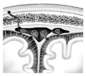
Figure 7-5
Frontal section through the superior sagittal sinus and longitudinal fissure.
Frontal section through the superior sagittal sinus and longitudinal fissure.
Such awesome responsibilities necessitate the brain be housed in the safe, bony structure of the cranial vault. It makes further good sense that this important organ should be wrapped in several layers of connective tissue meninges each of which has a special structure and function for nourishing, protecting and anchoring the brain.
The outermost or most superficial meningeal layer is the dura mater or "tough mother." This layer lives up to its name as it's a thick tough protective layer. The dura is anchored to and compartmentalizes the cranial vault. The compartmentalizing portion of the dura provides a system of venous "storm sewers," called venous sinuses, which return venous blood to the heart as fast as arterial blood is being delivered to the brain.
The dura has two identifiable layers. The outermost (periosteal) dura is really the periosteum of the inner side of the cranial vault bones and thus is firmly attached to the cranial vault. The inner (meningeal) dura is free to separate away from the outer dura and infold upon itself forming several incomplete walls or partitions. These partitions not only separate different parts of the brain (protecting the brain from excessive movement), but, by virtue of their double layer, form channels for venous blood drainage called venous sinuses. These venous sinuses receive large amounts of blood from all of the veins of the brain, eventually emptying into the internal jugular vein. (Figure 7-5)
The meningeal layer just deep to the dura mater is the arachnoid. This layer develops with the innermost meningeal layer the pia mater. During development, these two layers separate and form a sub-arachnoid space. When arachnoid is pulled away from pia mater, thin strands of arachnoid adhere to the pia. These arachnoid trabeculae give the arachnoid a spider web appearance and thus, its name. The subarachnoid space contains CSF. This fluid protects the brain and spinal cord forming a watery cushion. The pia mater, lying right next to the tissue of the brain and spinal cord, intimately follows all of the fissures, sulci and gyri of the CNS, and allows vessels to travel to and from the CNS. (Figure 7-5)