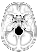
Figure 7-7
Floor of the cranial vault.
Floor of the cranial vault.
Shift your attention to the interior of the skull base with the brain removed. You can see that the floor of the cranial vault can be divided into three parts, forming the anterior, middle, and posterior cranial fossae.
The anterior cranial fossa, formed mainly by the frontal bone, is the roof of the orbits and supports the frontal lobes of the cerebrum. Its posterior margin is formed by the lesser wings of the sphenoid bone.
The middle cranial fossa is formed by the sphenoid and temporal bones, and supports the temporal lobes of the cerebrum. Many important structures pass through or are located in this fossa, including the optic nerves, the pituitary gland (hypophysis), the cavernous sinuses, the superior petrosal sinuses, the three divisions and ganglion of the V cranial or trigeminal nerve, and the internal carotid artery. (Figure 7-7)
The posterior cranial fossa is formed mainly by the occipital bone with a little help anteriorly from the temporal bone. This fossa houses and supports the cerebellum and the brain stem. The inferior end of the brain stem, the medulla, becomes continuous with the spinal cord as it passes through the large unpaired foramen magnum. Other structures found in the posterior cranial fossa include the transverse, inferior petrosal, and sigmoid sinuses. These latter two sinuses both pass through the jugular foramina as do cranial nerves IX, X, and XI. Cranial nerves VII and VIII leave the posterior cranial fossa through the internal acoustic meatus, while cranial nerve XII passes through the hypoglossal canal (found on either side of the foramen magnum). (Figure 7-7)