Figure 8-4
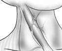
Superficial features of the posterior triangle.
Superficial features of the posterior triangle.
Begin in the posterior triangle of the neck. Remember, the anterior boundary of this triangle is the sternomastoid, and the posterior boundary is the trapezius. Identify these muscles and the external jugular vein that crosses the sternomastoid superficially and obliquely. (Figure 8-4)
Look at the floor of this triangle. From posterior to anterior the floor is made of the levator scapulae, scalenus posterior, medius and anterior.
If the sternomastoid and the posterior portion of the omohyoid have been cut and reflected, the trunks of the brachial plexus and the subclavian artery are seen as they pass between the middle and anterior scalene. The subclavian vein passes anterior to the scalenus anterior.
The inferior belly of the omohyoid has probably been removed, but it normally would lie just superficial to the latter three structures at the inferior part of the posterior triangle.
With the sternomastoid cut and reflected, you should be able to see several nerves on the floor of the triangle. From posterior to anterior, find cranial nerve XI, the spinal accessory nerve, lying on the levator scapulae; the phrenic nerve from C3,4,5 descends on the scalenus anterior on its way to the diaphragm; the cervical plexus C1-4 suppling sensory and motor nerves to the skin and muscles of the neck, including the anterior infrahyoid muscle group.