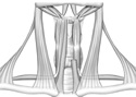
Figure 8-5
Anterior neck showing muscles of the anterior and submandibular triangles.
Anterior neck showing muscles of the anterior and submandibular triangles.
Let's move on to the anterior triangle of the neck beginning with the superficial structures. The muscular triangle will be our area of examination.
On either side of the midline there are four strap-like muscles, descending from the hyoid bone and thyroid cartilage. The infrahyoid muscles have names describing their attachments. The muscles effect the thyroid cartilage (larynx) and hyoid bone. They are active during swallowing, breathing and speaking.
The most superficial and medial of infrahyoid muscles are the paired sternohyoid muscles. They are located on either side of the midline. Just lateral to these, identify the superior portion of the omohyoid.
Lying deep to sternohyoid, (the sternohyoid has been cut on one side) you should be able to see the shorter sternothyroid and thyrohyoid. (Figure 8-5)
If you pull the two sternohyoid muscles apart, you should be able to see some of the cervical viscera. Identify the prominent "adam's apple" or laryngeal prominence (thyroid cartilage of the larynx ). Directly inferior to the adam's apple find the inferior end of the larynx, the cricoid cartilage, the trachea and the 1st tracheal ring and finally the isthmus of the thyroid gland. Gently pull the anterior cervical viscera (larynx, trachea, thyroid gland) laterally and expose the esophagus.
Superior and lateral to the omohyoid find the carotid triangle and its contents. Remember it is bounded superiorly by the posterior belly of the digastricus. Inferior to the posterior digastricus and slightly superior to the greater horn of the hyoid bone, you can see the hypoglossal nerve (cranial nerve XII) coursing towards the tongue. Cranial nerve XII supplies the tongue with motor innervation.
*Hint.: To feel the greater horn of the hyoid, steady the opposite horn with the same hand.
Inferior and deep to the hypoglossal nerve, find the internal and external carotid arteries. You have already identified branches of the external carotid in Exercise Six. Use those branches for orientation. Can you see the large internal jugular vein posterior to the carotid arteries? If you gently pull the internal jugular vein and the carotid arteries apart you will see the vagus nerve. In life, these three latter structures constitute a neurovascular bundle wrapped with a connective tissue "carotid sheath."
Proceed inferior to locate the bifurcation of the common carotid artery into the internal and external branches. The carotid sinus is a dilation at either the end of the common carotid or the beginning of the internal carotid artery. On the deep or medial side of the common carotid, see if you can find the superior end of the sympathetic trunk.
Examine the submandibular triangle, bounded by the posterior digastric, anterior digastric, and inferior margin of the body of mandible.. This anterior neck triangle contains salivary glands and most of the suprahyoid muscle group. Note how the two bellies of each digastric muscle are connected to each other by an intermediate tendon which in turn is attached to the hyoid bone. Find the slender stylohyoid muscle as it splits and goes on either side of the digastric tendon on its way to the hyoid bone. (Figure 8-5)
On one side of the cadaver, the large submandibular salivary gland should be visible. Part of this gland lies medial to the inferior border of the body of the mandible and superficial to the mylohyoid muscle. The mylohyoid muscle forms the floor of the mouth and part of this submandibular triangle's floor.
Find the hypoglossal nerve disappearing deep to the posterior border of this muscle. Can you see the facial artery and vein as they cross over the superficial aspect of the submandibular salivary gland? The last "subtriangle" in the anterior neck triangle is the sub-mental triangle. This small triangle lies between the midline of the neck and the anterior digastric. Identify the mylohyoid muscle. Just deep to the mylohyoid, find perpendicular running muscle fibers that belong to the paired geniohyoid.