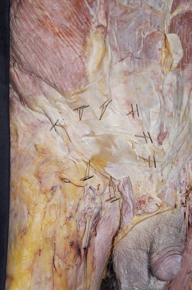
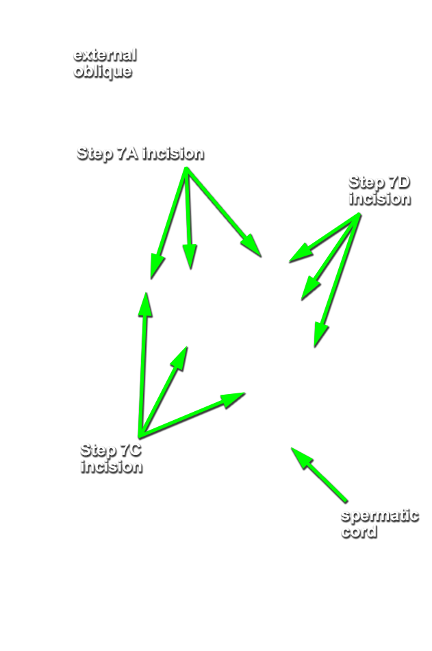
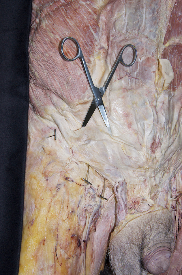
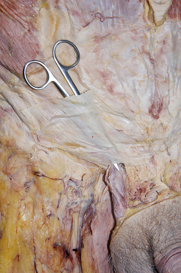
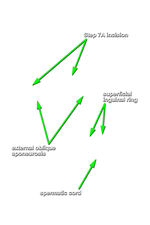
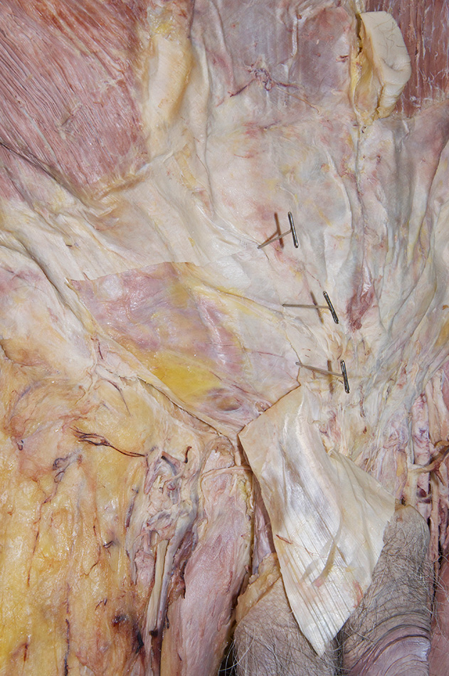
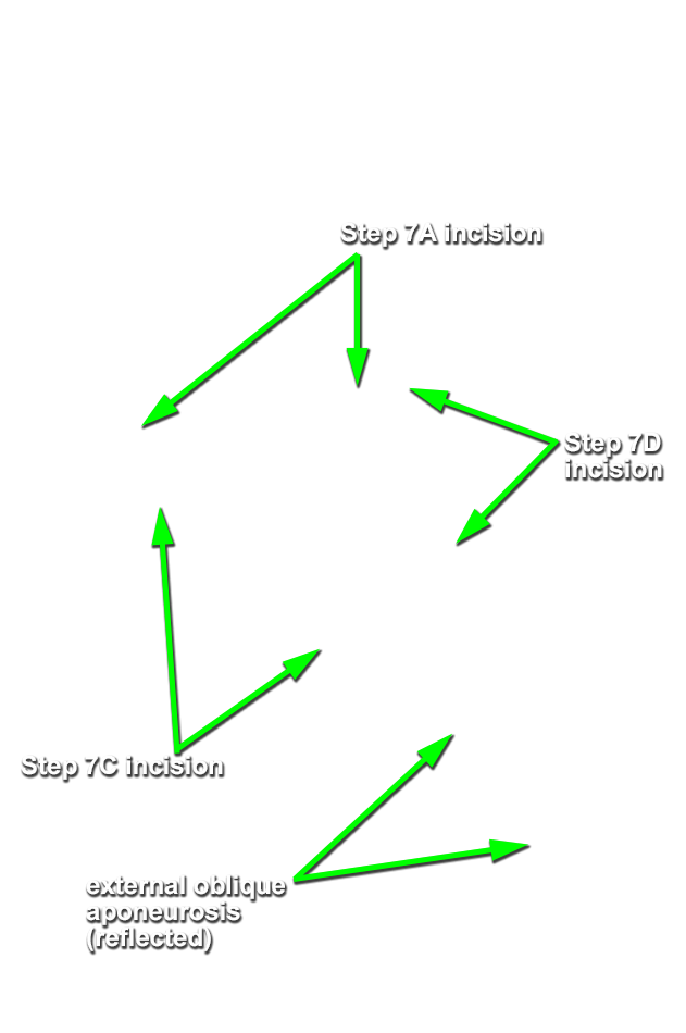
Dissect and identify the deep layers of the anterior abdominal wall on the right side of the cadaver. Before proceeding, carefully read step 7 and study the incision lines in the accompanying photograph for reflecting the aponeurosis of the external oblique muscle adjacent to the superficial inguinal ring.
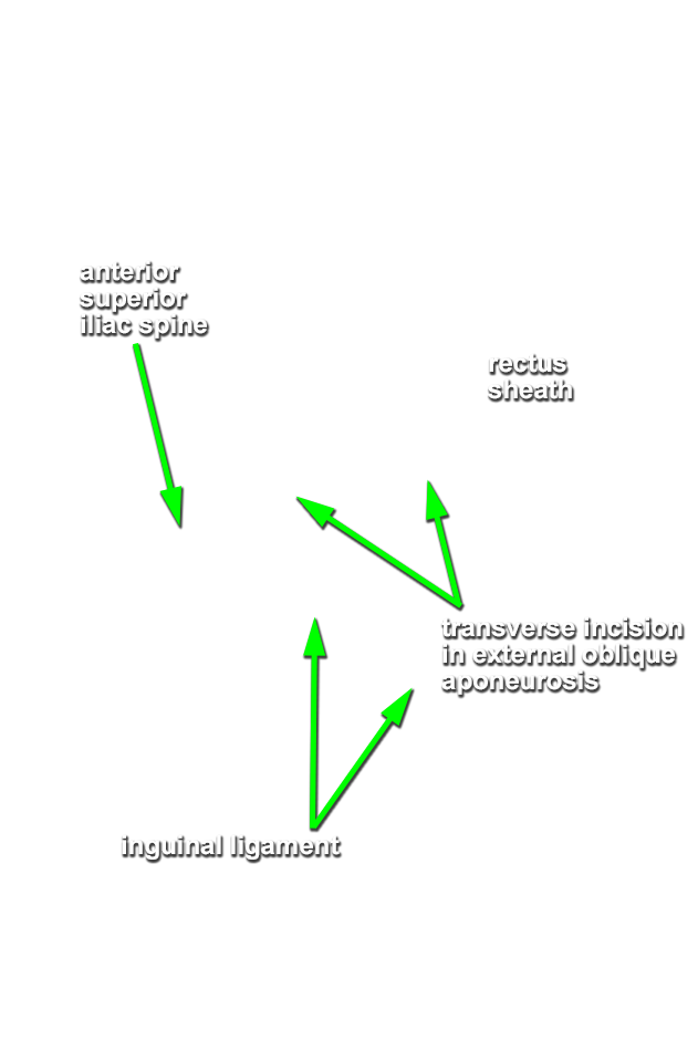
- Use scissors to make a transverse incision through the external oblique aponeurosis from the anterior superior iliac spine to the lateral edge of the rectus abdominis muscle.
- Use scissors (blunt dissect) and your fingers to separate the external oblique muscle from the underlying internal oblique muscle. Separate the two layers both at the superficial inguinal ring and the transverse incision through the aponeurosis of the external oblique muscle.
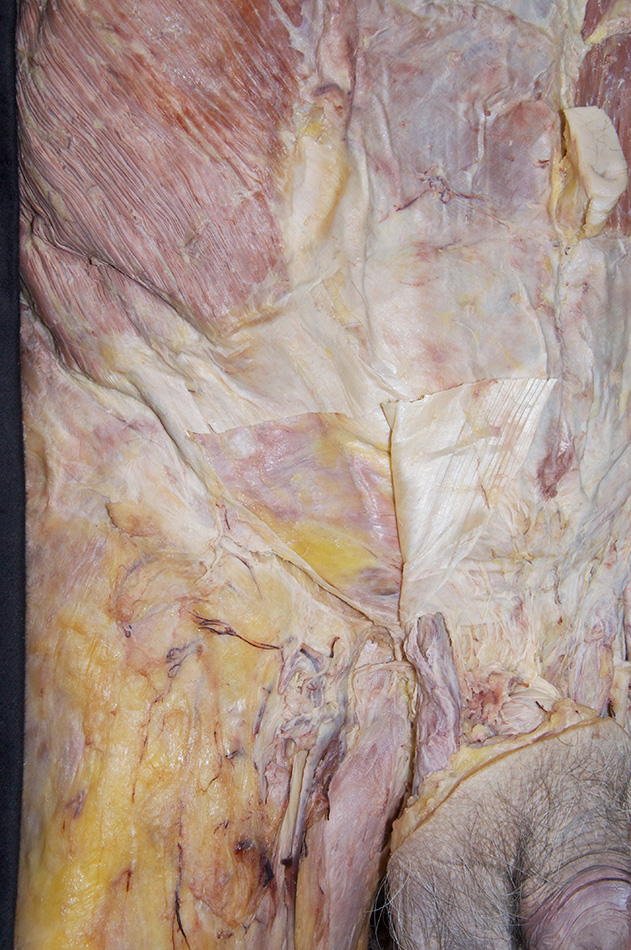
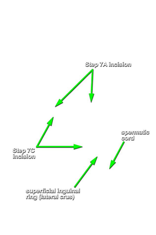
- Use scissors to make a vertical incision adjacent to the inguinal ligament. Carry the incision in the inferior direction from the anterior superior iliac spine torwards the superficial inguinal ring. Stop the incision approximately 1 cm superior to the lateral crus of the superficial inguinal ring. Be careful and do not cut the underlying muscle or the ilioinguinal nerve.
- Start at the level of the transverse incision (Step 7A incision) and use scissors to make a vertical incision through the external oblique aponeurosis adjacent to the rectus abdominis muscle. Carry the incision in the inferior direction stopping approximately 1 cm superior to the superficial inguinal ring.
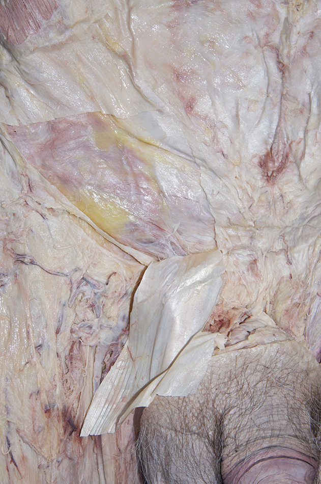
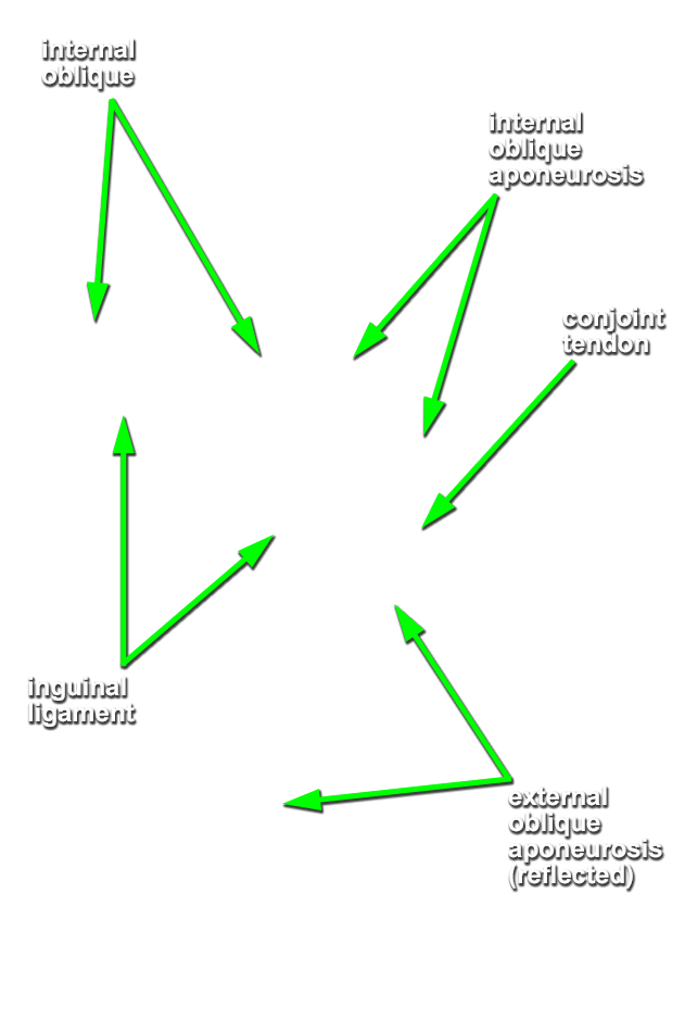
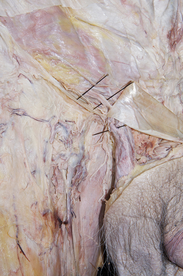
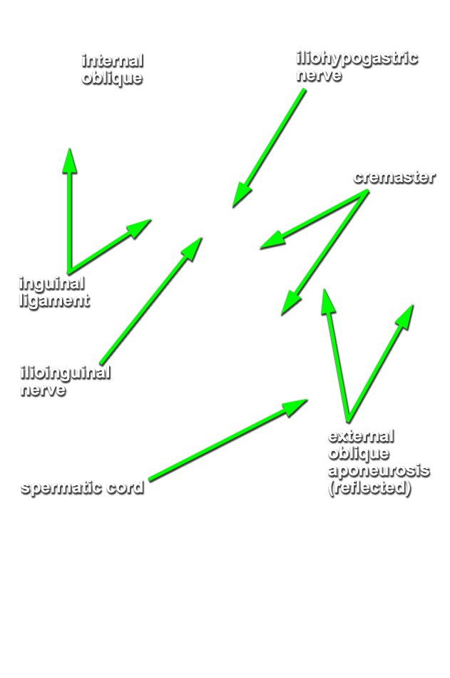
- Reflect (leave attached) the external oblique aponeurosis to expose the underlying muscle. Identify the internal oblique muscle, its aponeurosis, conjoint tendon and iliohypogastric nerve. (G 4.12A;N 253;Gl 13.13) Attempt to identify the cremaster muscle passing from the internal oblique to the spermatic cord.