Identify the coverings and structures of the spermatic cord and testes. Refer to an illustration of the anterior abdominal wall, spermatic cord and testes. (G 4.16;N 256;Gl 13.14) Note how each layer of the anterior abdominal wall contributes a covering of the spermatic cord and testes.
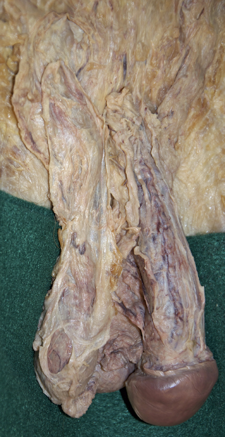
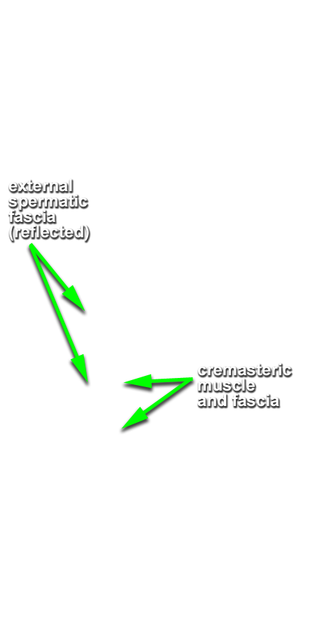
- Blunt dissect the testes free from the scrotal sacs. Identify theexternal spermatic fascia (G 4.16;N 365;Gl 13.14) covering the spermatic cord and testis. Use small scissors to make an incision in the external spermatic fascia and carefully reflect (do not completely remove) this layer from the spermatic cord and testis.
- Identify the cremaster muscle and fascia. (G 4.16;N 365;Gl 13.14) Use small scissors to incise this layer and reflect it from the spermatic cord and testis.
- Identify the internal spermatic fascia. (G 4.16;N 365;Gl 13.14) The cremaster, cremaster fascia and internal spermatic fascia may be fused. The outer surface is then the cremaster and the inner surface is the internal spermatic fascia.
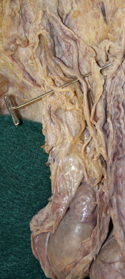
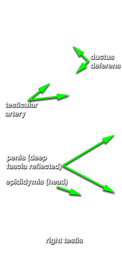
- Incise and reflect the internal spermatic fascia from the spermatic cord and testis. Identify the ductus deferens, testicular artery and testicular vein (pampiniform plexus). (G 4.19A;N 365;Gl 13.14) Attempt to identify the genitofemoral nerve and cremasteric and ductus deferens arteries.
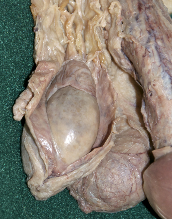
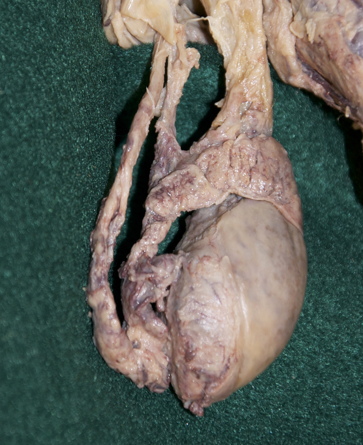
- Identify, incise and reflect the tunica vaginalis to expose the testis and epididymis. (G 4.19B;N 365;Gl 13.16) Trace (blunt dissect) the ductus deferens to the epididymis. Identify the tail, body and head of the epididymis. (G 4.20A;N 368;Gl 13.16) Use a scalpel to incise the capsule of the testis. Identify the tunica albuginea. (G 4.20B;N 368;Gl 13.16) Attempt to blunt dissect and identify one or more efferent ductules extending from the testis to the head of the epididymis.