Figure 2-13
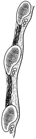
A parasagittal section through ribs 4, 5 and 6 of the thoracic wall showing vein, artery and nerve arrangement.
A parasagittal section through ribs 4, 5 and 6 of the thoracic wall showing vein, artery and nerve arrangement.
Begin on the outside of the anterior thorax. The pectoralis major and minor should be reflected.
In the fourth or fifth intercostal space near the lateral edge of the thorax, find and identify the external intercostal muscles. These muscles run inferiorly and medially like the front pockets of your jeans.
As you approach the sternum, note how the external intercostals seem to disappear, revealing the underlying internal intercostal muscles. The fibers of these muscles run at right angles to the external intercostals, i.e. inferiorly and laterally. The internal intercostals extend posteriorly in each intercostal space to the angles of each rib and then become membranous.
Deep to the internal intercostal muscles and just inferior to the rib above, in the costal groove, find the intercostal artery, nerve, and vein. These structures, from superior to inferior, are: vein, artery and nerve. (V.A.N.) Deep to these three structures you may be able to see the parietal pleura, which lines the thoracic wall. (Figure 2-13)
The thoracic wall has been cut laterally near the mid-axillary line and transversely along the
anterior attachment of the diaphragm. This dissection procedure has left the superior part of the
wall attached to the sternomastoid. The anterior wall can be lifted forward and upward like a trap
door to reveal the
transversus thoracis muscle
on the inner surface of the anterior wall of the chest. The fiber directions in the transversus
thoracis muscle allow it to aid in forced expiration. On the inner surface of the anterior wall,
the
internal thoracic artery
and
vein
can be seen running parallel to the sternum between the sternum and the transversus thoracis
muscle. Make an effort to find at least one
anterior intercostal artery
 as it comes off the internal thoracic artery.