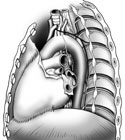
Figure 2-16
Left lateral view of the heart in the mediastinum showing the locations of major vessels and nerves.
Left lateral view of the heart in the mediastinum showing the locations of major vessels and nerves.
During the dissection procedure, the pericardial sac has been cut and opened, and the heart has been separated from its great vessels so it can be removed. However, it will be easier to identify general heart structures and some of the great vessels if the heart is left in its natural position.
In its natural position, the flat superior aspect of the heart is called the base. The more pointed inferior aspect of the heart, the point projecting to the left, is referred to as the apex of the heart. The largest part of the anterior surface and inferior border of the heart is made up of the right ventricle. The right border consists of the right atrium, while the left border and apex of the heart is formed by the left ventricle. The atria and ventricles can be visually separated by an atrioventricular (coronary) groove that encircles the heart in a superior transverse plane. This groove houses some of the coronary vessels. The ventricles are separated by anterior and posterior interventricular grooves. These grooves also house branches of the coronary vessels.
Continuing in the superior mediastinum, identify the great vessels entering and leaving the heart. Begin with the most prominent and anterior of these great vessels, the superior vena cava. This large vein empties into the right atrium and is formed superiorly by the joining of the left and right brachiocephalic veins. These two large veins are in turn formed by the joining of the internal jugular and subclavian veins.
With the heart still in place, look on the left side of the superior vena cava, and locate the short ascending aorta. Soon the ascending aorta will bend to the left and form the aortic arch. You will need to lift and reflect the left brachiocephalic vein to see the aortic arch and its major branches.
Identify major arteries as they arise from the aortic arch. From right to left they are: the brachiocephalic trunk (which soon splits into the right common carotid, and the right subclavian arteries), the left common carotid artery, and the left subclavian artery. The left vagus will cross the left side of the aortic arch between the left common carotid and the left subclavian artery.
Just to the left side of the aortic arch find and identify the pulmonary trunk which splits into two pulmonary arteries.
See if you can find the adult remnants of the fetal ductus arteriosus, the ligamentum arteriosum. In the fetus this was a small pulmonary vascular shunt. In the adult this has become a stout obliquely running ligament from the concavity of the aortic arch to the left pulmonary artery. (Figure 2-16)
Remove the heart from the thoracic cavity and examine it thoroughly. Hold it so it can be viewed in its natural position. Find the superior vena cava and the right atrium for orientation. Identify the inferior vena cava entering the inferior portion of the right atrium. Identify the right auricle of the right atrium.
Find the ascending aorta and pulmonary trunk. The aorta ascends from the left ventricle. The pulmonary trunk from the right ventricle. Just to the left of the pulmonary trunk are the left auricle and left atrium. Identify the left ventricle near the heart apex.
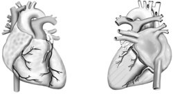
Next find and identify the coronary arteries. The prosectors should have removed most of the heart fat so you can see both the right and left coronary arteries. These arteries arise from either side of the ascending aorta and pass anteriorly, one on each side of the pulmonary trunk. Follow the right coronary artery as it leaves the ascending aorta. It should be in the coronary groove headed to the right border of the heart. Near the anterior right border of the heart, the right coronary artery sends a marginal branch winding toward the apex of the heart. The right coronary artery then proceeds around to the posterior aspect of the heart. When it reaches the posterior interventricular groove it gives off the posterior interventricular branch. (Figure 2-17)
Follow the left coronary artery from its origin on the left side of the ascending aorta. This artery is very short and soon divides under the cover of the left auricle near the pulmonary trunk into an anterior interventricular branch and the circumflex branch. The anterior interventricular branch lies in the anterior interventricular groove and descends towards the apex of the heart. The circumflex branch lies in the atrioventricular groove circling around the back side of the heart.
Most of the veins of the heart drain into the coronary sinus. This sinus lies in the posterior atrioventricular groove, empties into the right atrium and is formed from numerous tributaries.
Can you identify the tributaries of the coronary sinus? The great cardiac vein begins near the anterior apex of the heart and ascends in the anterior interventricular groove. It then turns sharply left and becomes continuous with the coronary sinus. The middle cardiac vein runs with the posterior interventricular artery, ascending in the posterior interventricular groove. It empties into the coronary sinus.
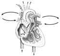
Finally, examine the interior of the heart by following the path of normal blood flow. (Figure 2-18)
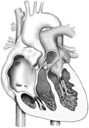
Begin the journey by finding the superior and inferior venae cavae. Open the right atrium and find the opening of the coronary sinus. It lies in the inferior part of this atrium. Just superior to the inferior vena cava find the adult remnants of the fetal interatrial shunt (foramen ovale), the fossa ovalis. (Figure 2-19)
The anterior atrial wall is rough and uneven due to the pectinate muscles attached to the surface. The posterior atrial wall is smooth. Both sinoatrial (SA) and atrioventricular (AV) nodes are located in the walls of the right atrium.
Move inferior and to the right to pass through the right atrioventricular valve (AV or tricuspid valve). This valve is composed of three valve cusps continuous at their bases. Papillary muscles arise from the floor of the right ventricle and have tendinous strands, chordae tendineae that come off their apices and attach to the margins of these valve flaps. (Figure 2-19)
Proceed through the right ventricle in a superior direction, passing through the pulmonary semilunar valve into the pulmonary trunk. The pulmonary valve is also composed of three valve cusps but does not possess any chordae tendineae or papillary muscles. Follow the pulmonary trunk from the right ventricle. It branches into left and right pulmonary arteries.
Identify the four pulmonary veins entering the left atrium. They carry oxygenated blood from the lungs into the heart. Gently use your probe to verify their entry into the left atrium.Now enter the left ventricle through the left atrioventricular valve (left AV, bicuspid or Mitral valve). Once inside the left ventricle, we can see the left AV valve is a two cusp valve, with chordae tendineae and papillary muscles like those found in the right ventricle. Note how thick the walls of the left ventricle are in comparison to the right ventricle.
Leave the left ventricle and pass through the aortic semilunar valve. This valve is very similar to the pulmonary semilunar valve, in that it has three valve flaps or cusps. Between the cusps and the aortic wall lie dilated pockets called aortic sinuses. These sinuses catch blood moving "backwards" and force the aortic semilunar valve flaps closed. There is always a good supply of arterial blood pooling in these sinuses - so it's no wonder the coronary arteries begin in two of these sinuses. Identify the openings for the coronary arteries found in the aortic sinuses.