Figure 2-15
Right lateral view of the heart in the mediastinum showing some important associated structures.
Right lateral view of the heart in the mediastinum showing some important associated structures.
Most of the parietal pleura has been removed to allow examination of many mediastinal structures. The phrenic nerves lay between the mediastinal parietal pleura and the fibrous pericardium. These nerves run longitudinally along the heart, near the lateral edge of the mediastinum to supply the diaphragm with motor and sensory innervation.
On the right side of the posterior mediastinum, locate the large azygous vein. You should be able to see it emptying into the superior vena cava.
On the left side of the posterior mediastinum, locate at least one posterior intercostal artery. You should see it arising from the descending or thoracic aorta.
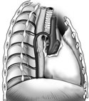
On either side of the posterior mediastinum find the sympathetic trunk. Follow it inferiorly as it crosses the heads of ribs 2-9. Identify the series of small swellings along the length of the trunk, the sympathetic ganglia. Sympathetic nerve processes link the ganglia. This linking is why these bilateral nerve trunks are sometimes called the sympathetic ganglionated chains. (Figure 2-15)
Find the
greater splanchnic nerve
 coming off the sympathetic trunk near the sixth intercostal space, on the left and right side. Can
you see the contributions this nerve receives from several of the sympathetic ganglia? The greater
splanchnic nerve carries preganglionic sympathetic fibers to the abdomen.
coming off the sympathetic trunk near the sixth intercostal space, on the left and right side. Can
you see the contributions this nerve receives from several of the sympathetic ganglia? The greater
splanchnic nerve carries preganglionic sympathetic fibers to the abdomen.
Turn your attention to the area above the heart, the superior mediastinum. Begin on the right side of the mediastinum by again identifying the azygous vein. Remember it empties into the superior vena cava. Still on the right side and just posterior to the superior vena cava, find the trachea. On the lateral side of the trachea, locate the right vagus nerve. At the bifurcation of the trachea, near the fifth intercostal space and posterior to the root of the lung, the vagus nerve leaves the trachea and comes to lie on the lateral edge of the esophagus. Posterior to the trachea and to the right, find the esophagus. (Figure 2-15)