Figure 2-14
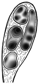
The arrangement of arteries, veins and bronchi in the hilus of the left lung.
The arrangement of arteries, veins and bronchi in the hilus of the left lung.
Roots of the lungs are cut allowing them to be lifted from the thoracic cavity revealing other important thoracic structures. Examining the lungs, you will see that the right lung has three lobes. The left lung has only two. Examine the medial sides and find the cut roots of the lungs. Try to identify the pulmonary veins, pulmonary arteries and the bronchi. Generally, the bronchus lies posterior and has cartilaginous rings (the pulmonary artery superior and the pulmonary vein inferior). (Figure 2-14)
With the lungs removed, identify and examine the extent of the
diaphragm.
Take time to appreciate the domed structure of this organ. Take time to Identify the nonmuscular
central tendon
 .
.