
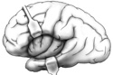
Figure 7-8
Hemisected brain (right half).
Hemisected brain (right half).
Review Figure 7-2. Keep in mind the specimen you are using may have the filmy
arachnoid
 around it. Identify the two large
right and left cerebral hemispheres. The two cerebral hemispheres are separated from one another by a deep mid-sagittal
longitudinal fissure
.
The floor of this fissure is the
corpus callosum
which is made of nerve fibers that connect the two cerebral hemispheres.
around it. Identify the two large
right and left cerebral hemispheres. The two cerebral hemispheres are separated from one another by a deep mid-sagittal
longitudinal fissure
.
The floor of this fissure is the
corpus callosum
which is made of nerve fibers that connect the two cerebral hemispheres.
At the posterior inferior surface of the cerebrum identify the cerebellum also composed of two hemispheres. Find the brain stem on the base of the cerebrum (Figure 7-3). Can you see any of the cranial nerves as they emerge from the brain stem?
Look at the cerebral and cerebellar hemispheres and the brain stem using a hemisected brain (Figure 7-8).
Begin by identifying the various sulci, notches and fissures that separate the lobes of the cerebrum and then identify the lobes themselves. Some anatomists include another area of the cerebral cortex, the insula as a separate lobe. The insula is very hard to see as it is buried beneath the other lobes of the brain. (Figure 7-9)