 . This sulcus runs in a posterosuperior direction beginning from a prominent cleft in the anteroinferior part of the cerebrum, the sylvian fossa. (Figure 7-2)
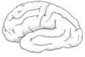
Our journey through the cerebrum begins with identification of several important sulci. First, on the lateral surface of the cerebral cortex, find the
lateral sulcus
. This sulcus runs in a posterosuperior direction beginning from a prominent cleft in the anteroinferior part of the cerebrum, the sylvian fossa. (Figure 7-2)
The
central sulcus
begins as a notch between the lateral and medial hemispherical surfaces and runs anteroinferior towards the lateral sulcus, but it usually does not reach the lateral sulcus. The central sulcus separates the
precentral gyrus
(primary motor cortex) from the
postcentral gyrus
(primary somatic sensory cortex). (Figure 7-2)
On the posterior medial aspect of the cerebral hemisphere surface of a hemisected brian, you can easily see the
parieto-occipital sulcus
and the
calcarine sulcus
. These two form a "Y" lying on its side (the calcarine sulcus forming the stem and the inferior wing of the Y, while the parieto-occipital sulcus forms the superior wing). The stem of the "Y" begins just inferior to the posterior part of the corpus callosum. (Figure 7-8) Our last boundary maker is the
pre-occipital notch
. This notch is located on the posterior and inferolateral hemisphere surface. (Figure 7-10)
Using these sulci and notches identify the four lobes of the cerebral hemisphere. The frontal lobe extends from the anterior tip of the cerebrum to the central sulcus posteriorly and the lateral sulcus inferiorly.
Note the most posterior gyrus of the frontal lobe, the
precentral gyrus
.
Remember this area is the primary motor area of the cerebral cortex.
In order to visualize the remaining three lobes, we need to draw two imaginary lines on the lateral hemispherical surface. The first line extends from the parieto-occipital sulcus, where it passes from the lateral to the medial hemispheric surface, to the preoccipital notch. The second line runs from the posterior end of the lateral sulcus in a posterior direction, to the point where it intersects the first line. (Figure 7-10)
The
parietal lobe
is bounded anteriorly by the central sulcus, posteriorly by the imaginary line uniting the parieto-occipital sulcus to the preoccipital notch, and inferiorly by the lateral sulcus and its imaginary posterior extension. Once again, note the most anterior gyrus of the parietal lobe, the
postcentral gyrus
.
This is the primary sensory area of the cerebral cortex. The
occipital lobe
lies posterior to the first imaginary line, posterior to the parietal lobe and extends to the posterior tip of the cerebrum.
The temporal lobe lies anterior to the occipital lobe and inferior to the lateral sulcus and thus inferior to both the frontal and parietal lobes.